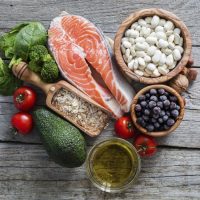
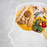
About Your Brain
First, put your two fists together, fingers touching. This is the size of your brain. It weighs about 3 pounds. Together, the brainstem and midbrain control breathing and heartbeat, sex drive, consciousness, and body temperature. The inner structures are surrounded by the wrinkly cortex - the thinking, processing part.
Albert Pike
"To work with the hands or brain, according to our requirements and our capacities, to do that which lies before us to do, is more honorable than rank and title."
Contemplating your own brain?
Here is a basic tour:
First, put your two fists together, fingers touching. This is the size of your brain. It weighs about 3 pounds. Now, imagine it is made of grayish pudding. It is scintillating, literally popping, with electrical signals from the 100 billion neurons it contains. For each neuron, the cell that does the “work” of thinking, there are about 9 support cells, called glia, which help each neuron to do its job.
Your brain sits on a stalk (your wrists) called the brainstem, which extends deep into the brain, connecting with midbrain structures. Together, the brainstem and midbrain control many functions – like breathing and heartbeat, sex drive, consciousness, and body temperature. The inner structures are surrounded by the wrinkly cortex (your fingers) - the thinking, processing part.
Now, imagine a ring-like cradle of blood vessels at the base of your brain, climbing like tendrils up the sides, and penetrating inward. These blood vessels taper until they become capillaries, supplying each brain cell with oxygen and nourishment, carrying away waste, and keeping the brain cool.
Projecting from the front of your brain, you see two thick stalks made of neural tissue, attached to the back of your eyeballs.
Got the picture? Read on!
Parts of the Brain
All the parts of the brain work together, but each part has its own special properties.

The Cerebellum
(pictured in red)
Learning, Movement and Social Skills
When you play the piano or hit a tennis ball you are activating the cerebellum. It coordinates your movement. As you walk, sit or move, constant communication between the limbs, spine and cerebellum help you maintain your balance with small imperceptible adjustments.
New research shows that the cerebellum is important in learning and development of social skills.
The Cerebrum
Planning, Thinking and Remembering
The cerebrum is the larger part of the brain that you might think of first, when contemplating your own brain. It holds your memories, allows you to plan, enables you to imagine and think. It allows you to recognize friends, read books, and play games.
The cerebrum is split into two halves (hemispheres) which communicate with each other through a thick bundle of nerve fibers called the corpus collosum.
Parts of the Cerebrum: Learning About Lobes
Each cerebral hemisphere is divided into sections, or lobes.
Each lobe specializes
in different functions. However, the brain is “plastic” and adapts itself to
you as an individual. People who spend more time developing physical skills may
have more brain area devoted to certain movements, while those who have
developed musical skills may have more brain area devoted to their skill set.
Frontal Lobes: Thinking and Planning
Frontal lobe shown in yellow
Here are some functions of the frontal lobes:
Thinking and Reasoning
Here are some functions of the frontal lobes:
Thinking and Reasoning
When you plan a schedule or imagine the future, the two frontal lobes do much of the work. One of the ways the frontal lobes seem to do these things is by acting as short-term storage sites, allowing one idea to be kept in mind while other ideas are considered.
Controlling Movement
At the rear of each frontal lobe is a motor area which helps us to control our movements.
Putting Thoughts Into Words
A place on the left frontal lobe called Broca's area helps us express our thoughts in words.
Parietal Lobes: Making Sense of the Senses

Parietal lobe shown in blue
When you enjoy a good meal you experience the taste, aroma, and texture of the food. The parietal lobes are at work helping to process information from your senses.The forward parts of these lobes are known as the primary sensory areas (7) which receive information about temperrature, taste, touch, and movement from the rest of the body.
These bits of information are then combined to form a complete picture of the experience.
Reading and math are also processed in each parietal lobe.
Occipital Lobes: Like Eyes in the Back of Your Head
![]()
The occipital lobe is shown here in a lovely lavendar
As you look at the words and pictures on this page, two areas at the back of the brain, called the occipital lobes are at work. The optic nerve attaches to the back of the eye (remember the wiggly eyes at the top of the page?)
Temporal Lobes: Processing What You Hear

Temporal lobe shown in green
At the top of each temporal lobe is an area responsible for receiving information from the ears.
The underside of each temporal lobe plays a crucial role in forming and retrieving memories, including those associated with music.
Other parts of this lobe seem to link memories and sensations of taste, sound, sight, and touch.
The Cerebral Cortex
Coating the surface of the cerebrum and the cerebellum is a layer called the cortex. Most of the actual information processing in the brain takes place in the cerebral cortex.
The cortex contains 6 layers of cells that work to process information. Folding the cortex increases the amount of gray matter that can fit into your skull, and the quantity of information that can be processed in the available space (aren’t you glad it’s folded? Otherwise your brain would be enormous!
If you unfolded the cortex, you would find it’s about the size of a pillowcase. (Think about that next time you put your head on a pillow!)
The cortex is gray because nerves in this area lack the shiny white insulation called myelin.
Beneath the cortex are neurons coated with myelin. This myelinated brain tissue is known appropriately as “white matter.”
The Inner Brain: Basics of Life

Limbic System: Processing Emotions
We often say that our emotions run deep, and they are processed deep within the brain in an area known as the limbic system. The limbic system is so named for its ring-like appearance. It is closely linked to the memory processing area known as the hippocampus.
Hypothalamus: Coordinating Body and Emotions
The hypothalamus is about the size of a pearl, but directs a multitude of important functions. It wakes you up in the morning, and gets the adrenaline flowing during a test or job interview. The hypothalamus is also an important emotional center, controlling the molecules that make you feel exhilarated, angry, or surprised.
Thalamus: Information Relay Station
Near the hypothalamus lies the thalamus, a major clearinghouse for information going to and from the spinal cord and the cerebrum. It acts to help the brain know what is going on in our outside world, and then send instructions out to the body to move and respond as needed.
Hippocampus: Emotion and Memory
The hippocampus is essential for memory processing. This tiny area helps prioritize what we need and discard unnecessary detail – a process of refining memory that we perform constantly during the day. During sleep, the brain shifts its function to continue deeper processing and solidifying of memory.
To store memory, the hippocampus sends memories out to the appropriate part of the cerebral hemisphere for long-term storage. The memory is stored in an interesting way: instead of storing an entire event - such as patting a puppy, each retrieves the information when necessary.
As mentioned earlier, memory (hippocampus) and emotion (limbic) areas are closely linked. You have probably noticed that emotionally charged experiences are hard to forget.
Basal Ganglia: Moving Smoothly
The basal ganglia are clusters of nerve cells surrounding the thalamus. They are responsible for getting a movement started, and helping it to flow in a purposeful way.
Neurons

Neurons consist of three parts:
There are different types of neurons in different areas, but here are some basic features they share:
The cell body contains the nucleus, where most of the molecules that the neuron needs to survive and function are manufactured.
Dendrites extend out from the cell body like the branches of a tree and receive messages from other nerve cells. Signals then pass from the dendrites through the cell body and travel away from the cell body down an axon to another neuron. Some axons are wrapped with a fatty insulation called myelin which allows the signal to travel faster.

Synapses change as we learn from our experiences. The brain forms new synapses when we are learning, and it also prunes away synapses we don't need. In this way, our brain is constantly adapting to reflect our lives.
The of messenger chemicals, called neurotransmitters, are released into the space between the neurons, called the synapse.
The receptors "catch" the neurotransmitter molecule, triggering a new electrical signal that will travel to the next neuron.
Some Key Neurotransmitters at Work
There are dozens of neurotransmitters but here is a description of some of the key neurotransmitters:
-
Acetylcholine: Movement and Memory
Alzheimer’s disease, which initially affects memory formation, is associated with a shortage of acetylcholine.
-
GABA: Calming, Controlling
neurotransmitter because it has a dampening effect, causing cells to be less excitable.
-
Serotonin: Mood
-
Dopamine: Mood, Movement, Motivation and Learning
Glia - Support Cells for Neurons
There are about 9 support cells for every neuron. Glia are fascinating because they support neurons in so many ways - nourishment, protection, insulation. Scientists are learning more about this "behind the scenes" brain cell.
One type of glial cell wraps around the neuron, forming a fatty insulation called myelin.
Myelin helps nerve signals travel fast – up to 400 miles per hour!
Another type of glial cell helps to nourish neurons by helping to process nutrients.
This overview of the brain only touches the surface, so to speak!

